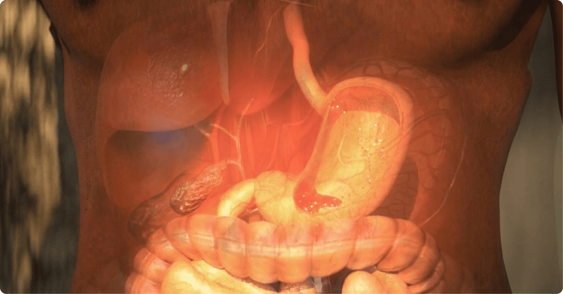
The immune system is one of the body’s most sophisticated networks, responsible for defending us against a constant stream of threats. Composed of specialised cells, tissues, and organs, it works together to identify and neutralise pathogens as well as the body’s own cells when they turn abnormal.
Autoimmune diseases, such as rheumatoid arthritis, occur when this distinction breaks down. The immune system mistakes the body’s own tissue for a threat and attacks it, driving chronic inflammation and progressive damage.
Explaining how antibodies lock onto antigens or how T cells spot infected cells can feel like describing an invisible world, especially when talking to non-scientific audiences. The three-dimensional representation of an antibody binding to its antigen or a T cell recognising an infected cell makes these microscopic processes easy to understand for both expert and lay audiences, enhancing knowledge retention.
Are you interested in modular content and omnichannel materials?
The Key Organs of the Immune System
- Bone Marrow: The soft tissue inside our bones and the birthplace of all immune cells. Through a process called haematopoiesis, it produces the white blood cells (leukocytes) that form the backbone of the body’s defence.
- Thymus: Located behind the breastbone, the thymus is where T cells (T lymphocytes) mature and are “educated” to recognise the difference between the body’s own cells and foreign threats.
- Lymph Nodes: Small, bean-shaped structures distributed throughout the body. They filter lymph and act as meeting points where immune cells encounter antigens and launch tailored adaptive responses.
- Spleen: Positioned in the upper left abdomen, the spleen filters the blood, removes worn-out red blood cells and captured pathogens, and houses a large reservoir of immune cells ready to respond.
- Lymphatic Vessels: A body-wide transport network that carries lymph, along with immune cells and antigens, between tissues and lymph nodes, keeping the system connected and responsive.
- Mucosal and Skin Barriers: The first line of defence. Beyond acting as physical barriers, the skin and the mucosal linings of the gut and airways contain localised immune tissue that intercepts threats at the point of entry.
Get this free Guide with
5 key steps for The Digital Transformation
of Scientific Communication
Immune Cells and Their Functions
The immune system relies on a diverse cast of cells, each with a specialised role. Let’s explore some of the most important players and what they do:
- Antibodies and Cytokines: Antibodies (immunoglobulins) bind to antigens with remarkable specificity, neutralising or tagging them for destruction. Cytokines are the signalling molecules that orchestrate communication across the entire immune network.
- Macrophages and Neutrophils: The frontline phagocytes of the innate immune system. They engulf and destroy pathogens on contact and raise the alarm that recruits other cells to the site of infection.
- Dendritic Cells: Expert messengers that capture pathogens, process them, and present their fragments to T cells. They bridge the fast innate response and the precise adaptive one.
- T Lymphocytes (T cells): Helper T cells (CD4+) coordinate and amplify the immune response, while cytotoxic T cells (CD8+) seek out and destroy infected or abnormal cells directly.
- B Lymphocytes (B cells): The antibody factories. When activated, they differentiate into plasma cells that produce antibodies targeted to a specific antigen, and into memory cells that remember the threat for next time.
- Natural Killer (NK) Cells: Rapid-response patrollers that identify and eliminate virus-infected and cancerous cells without needing prior exposure.
Antibodies, Antigens, and Autoimmune Disease
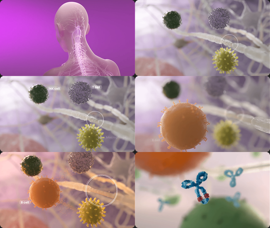
Utilising advanced 3D visualisation techniques makes it possible to create scientifically accurate, visually striking videos, such as the moment an antibody locks onto its target antigen.
In the finely tuned choreography of the immune system, recognition is everything. Antigens are the molecular markers found on the surface of pathogens: the flags that identify a threat. In response, B cells and plasma cells produce antibodies that bind to these antigens like a key in a lock, neutralising the invader or tagging it for destruction, while T cells coordinate the wider response and eliminate infected cells.
Underpinning all of it is a single, vital ability: distinguishing self from non-self, so the system attacks danger and leaves healthy tissue untouched.
In autoimmune disease, that distinction breaks down. The immune system mistakes the body’s own cells for a threat and turns its weaponry inward. In rheumatoid arthritis, for example, immune cells and antibodies target the synovium — the lining of the joints — triggering chronic inflammation, pain, and progressive joint damage. Genetics, environmental triggers, and immune dysregulation all contribute to this loss of tolerance.
3D animation allows you to present these intricate processes in a visually engaging and scientifically precise manner. The three-dimensional depiction of antibody–antigen binding, T cell activation, or a cytokine cascade turns abstract, microscopic events into reliable educational tools for researchers, healthcare professionals, and patients alike.
The same recognition machinery is now harnessed in modern immunotherapies, from monoclonal antibodies to checkpoint inhibitors, explaining it through a therapy’s mechanism of action video.
How can 3DforScience help you?
3DforScience is a scientific communication company specialising in 3D animation and visual content for the life science sector. We are a passionate creative team of scientists, marketers, artists, and animators with a shared aim: to help you improve your scientific communication with innovative and unique creative solutions.
We can help boost your scientific communication capabilities across immunology, molecular biology, and biotechnology by designing tailored creative solutions such as 3D animation videos and illustrations, turning complex mechanisms into content your audiences actually understand and remember.
Would you like to bring your scientific messaging to life? Contact us. We would be pleased to provide you with a unique creation.
- What types of immunology treatments can be visualised with 3D animation?
From monoclonal antibodies and checkpoint inhibitors to cell therapies like CAR-T, we can bring any immunological intervention to life. - Who benefits from 3DforScience’s medical animations?
Everyone wins: pharma teams get clearer communication tools, while HCPs and patients gain a much easier way to grasp complex science. - How does 3D animation help explain immunological Mechanisms of Action (MoA)?
It turns abstract molecular interactions into clear, step-by-step visual stories that make the “how” and “why” instantly understandable. - Can these immunological animations be adapted for VR or medical congresses?
Absolutely, our assets are built to be flexible, working seamlessly in immersive VR experiences or as standout visuals at major medical congresses. - How long does it take to produce a 3D animation for an immunological MoA?
Timelines vary based on complexity and the length of the video, but we typically aim for 6-8 weeks to ensure both scientific accuracy and high-quality storytelling.