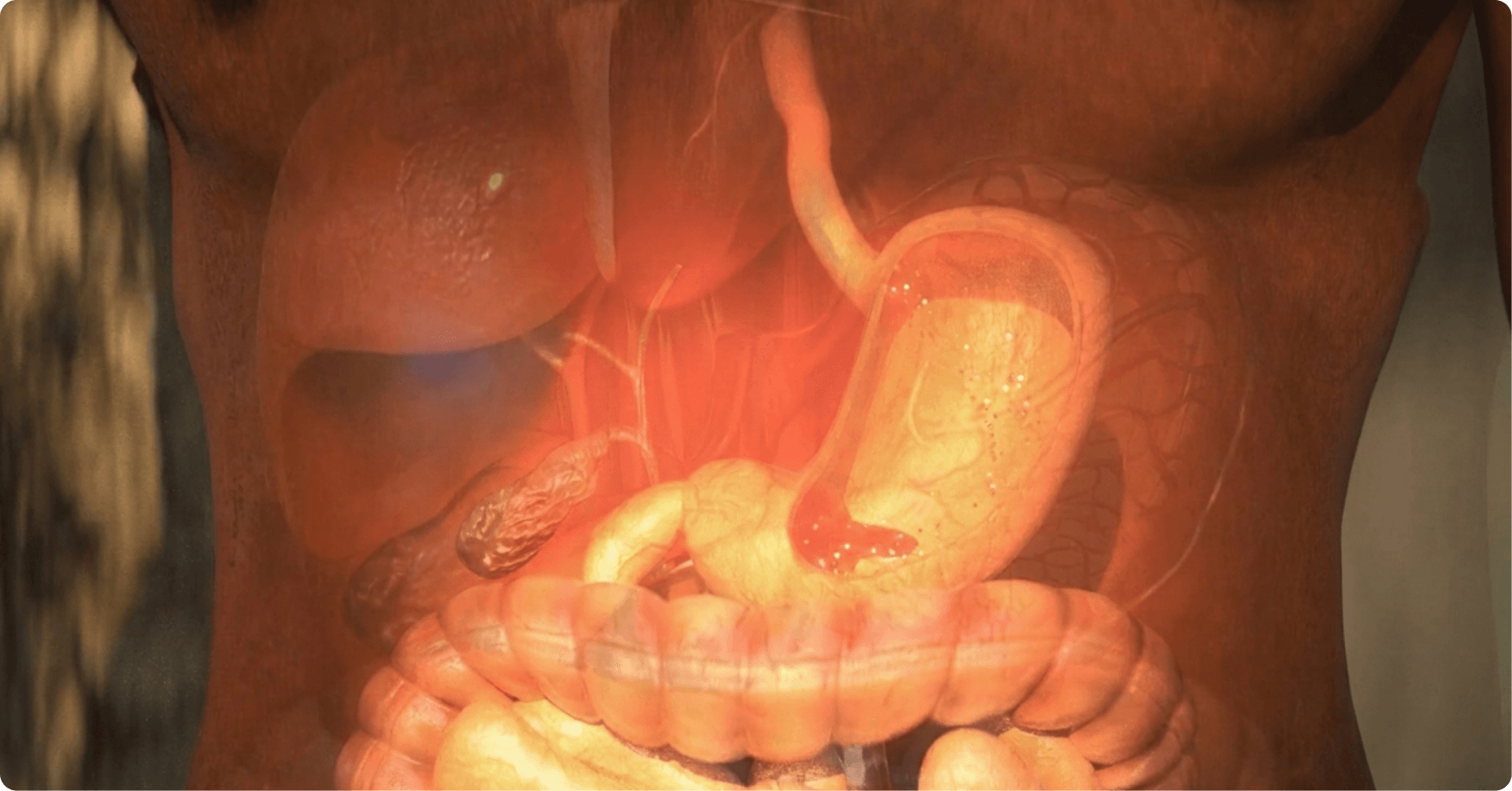
Of all the organ systems in the human body, the digestive system is the one your audience has almost certainly never seen at work.
It runs some nine metres from mouth to colon, folded and coiled into the abdomen, and everything it does happens in the dark: churning, secreting, absorbing, defending, and holding a conversation with the brain and with trillions of microorganisms at the same time.
For pharmaceutical, biotech and medical device companies working in gastroenterology, that invisibility is the communication problem. A mechanism of action that plays out on the surface of a colonic epithelial cell, or a device that travels the length of the small intestine, cannot be filmed. It can be animated.
This article walks through how 3D medical animation is used across gastroenterology, from the anatomy of the gut to the types of video that GI companies commission most often, using our own work in the field as reference.
Are you interested in modular content and omnichannel materials?
The digestive system, visualised
The gastrointestinal tract is a continuous tube with a very different job at each stage.
In the mouth and oesophagus, food is broken down mechanically and pushed along by waves of muscular contraction known as peristalsis.
In the stomach, parietal cells secrete hydrochloric acid while the mucosa protects itself with a layer of mucus and bicarbonate, a balance that fails in gastritis, peptic ulcers and reflux.
In the small intestine, the wall folds into villi and microvilli, creating an absorptive surface many times larger than the skin, where enzymes from the pancreas and bile from the liver and gallbladder complete digestion and nutrients cross into the bloodstream.
In the colon, water is reclaimed and the gut microbiome, trillions of bacteria and other microorganisms, ferments what remains, producing short-chain fatty acids that feed the colonic epithelium and modulate the immune system.
Two further layers make gastroenterology such a rich subject for animation:
- The first is the gut barrier: a single layer of epithelial cells, renewed every few days and sealed by tight junctions, that separates the contents of the gut from the largest concentration of immune tissue in the body.
- The second is the enteric nervous system, hundreds of millions of neurons embedded in the gut wall and in constant dialogue with the central nervous system through the brain-gut axis. Stress, inflammation, motility and mood all meet here, which is why so many GI conditions are as much about signalling as they are about structure.
None of this can be seen through an endoscope. All of it can be shown with a 3D camera that moves freely from the lumen to the lamina propria, from the whole organ to a single receptor.
Get this free Guide with
5 key steps for The Digital Transformation
of Scientific Communication
Types of medical animation in gastroenterology
Every format we describe in our overview of the types of medical animation has a gastroenterology version. What changes is the science inside it.
Mechanism of disease animation
Before an audience can understand a treatment, it needs to understand what has gone wrong, and in the gut the pathology is rarely visible from the outside.
That is why most GI projects begin with a mechanism of disease (MoD) animation.
In inflammatory bowel disease, that means showing how the barrier becomes leaky, how immune cells infiltrate the mucosa and how the cycle of inflammation sustains itself in Crohn’s disease and ulcerative colitis.
In gastro-oesophageal reflux disease it means the lower oesophageal sphincter and the damage acid does to the oesophageal lining.
In coeliac disease it is the immune reaction to gluten that flattens the villi.
In metabolic dysfunction-associated steatohepatitis (MASH) it is fat accumulating inside hepatocytes, followed by inflammation and fibrosis.
Each of these stories has a scale and a timeline that live footage cannot capture and that a 3D animation can compress into ninety seconds.
Mechanism of action animation
Gastroenterology is one of the most active therapeutic areas in pharma, and its pipeline is molecular.
Proton pump inhibitors block the H+/K+ ATPase in the parietal cell.
Biologics for IBD neutralise TNF, block the α4β7 integrin that guides lymphocytes to the gut, or intercept interleukin-23, while small molecules such as JAK inhibitors and S1P receptor modulators act inside the cell or on lymphocyte trafficking.
Microbiome therapeutics work by restoring bacterial communities rather than hitting a single target.
A mechanism of action (MoA) animation gives each of these a clear visual identity: the receptor, the ligand, the moment of binding and the downstream effect on the tissue. For medical affairs and marketing teams it is the single most reused asset in a GI launch, appearing in congress booths, MSL decks, websites and HCP training.
Cellular and molecular animation
Some gastroenterology stories are best told at the scale of a single cell.
An enteroendocrine L cell releasing GLP-1 in response to nutrients, a goblet cell secreting mucus, a bacterium producing butyrate at the epithelial surface or a tight junction opening under inflammatory signalling are all subjects for cellular and molecular animation.
They are used in scientific education, in investor and partnering material for biotech companies, and as building blocks that can be reused across an entire campaign.
Medical device animation
The GI tract is also one of the busiest fields for devices.
Endoscopes and capsule endoscopy systems, biliary and oesophageal stents, intragastric balloons, motility and pH monitoring probes and diagnostic breath tests all need to be explained to clinicians, purchasers and sometimes patients.
Medical device animation shows how a device is positioned, how it works once in place and what the clinician sees, without the constraints of filming inside a living patient. It doubles as a training tool for sales teams and for the healthcare professionals who will use the device.
Surgical and procedural animation
Colonoscopy, endoscopic mucosal resection, ERCP, fundoplication, bariatric procedures such as sleeve gastrectomy and gastric bypass, and liver and colorectal surgery are all routinely animated, either as step-by-step training for surgeons and endoscopists or as simplified versions that help patients understand what will happen to them.
Combining real surgical footage with 3D reconstruction is a particularly effective approach here, because the animation can reveal the anatomy the camera cannot reach.
Patient education animation
Many GI conditions are chronic, invisible and stigmatised, and adherence depends on understanding.
Patient education animation translates the biology of IBS, IBD, reflux or liver disease into plain, empathetic visuals, and is increasingly delivered in immersive formats.
Our project with the creative agency Grow is a good example: a 3D animated video with a virtual reality adaptation that explains how stress acts on the brain-gut axis, from cortisol release to changes in the microbiome and the symptoms patients feel, designed to help people with stress-related digestive disorders understand and manage their condition. Read our Grow case study
Pharmaceutical technology and platform animation
Finally, the gut itself is a drug delivery challenge.
Enteric coatings, modified-release formulations, colon-targeted delivery, oral biologics and microbiome engineering platforms all need an explanation of where in the tract the technology acts and why that matters.
Platform animation makes the value of a formulation or a delivery system visible to partners and investors long before clinical data are available.

Gastroenterology at 3DforScience
Gastroenterology is one of the specialties in which our studio works most regularly, alongside oncology, immunology and dermatology.
Our in-house scientists and pharmacists develop the scientific storyline for each project, checking every structure, pathway and claim against the literature before our designers and animators begin, so that the finished video is accurate enough for medical review and clear enough for a patient.
We mix real footage with 3D elements where it helps, adapt the same assets for congress screens, web, social media and VR, and work directly with pharma, biotech, device and agency teams rather than through third parties.
If your product acts somewhere between the oesophagus and the colon, we can show your audience exactly where and how.